Ever wonder why your doctor orders a resting heart test and then asks you to run on a treadmill? It's because your heart behaves differently when you're lounging in a clinic than when you're pushing your limits. While a standard heartbeat recording is great for a snapshot, some heart problems only show up when the engine is actually running. Understanding the difference between these tests can take the mystery out of your next cardiology appointment and help you advocate for the right diagnostic path.
| Feature | Resting ECG | Stress Test |
|---|---|---|
| Purpose | Baseline electrical rhythm | Heart response to exertion |
| Duration | 3-5 minutes | 10-60 minutes |
| Activity | Lying still | Treadmill or chemical agents |
| Key Detection | Arrhythmias, past heart attacks | Silent ischemia, CAD |
The Basics of the Resting ECG
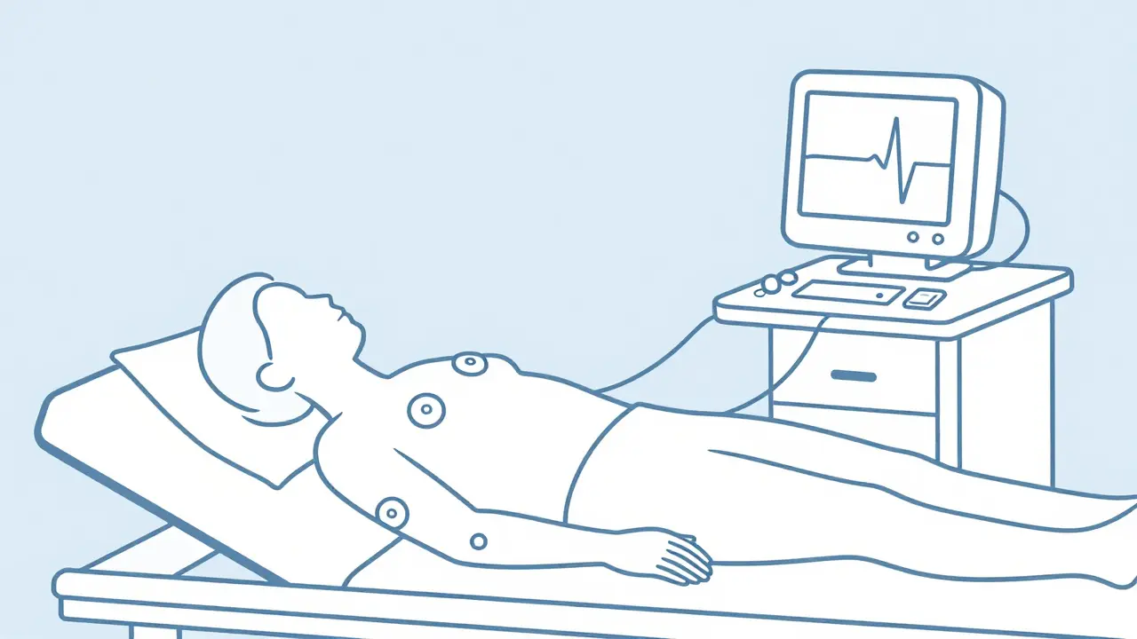
At its core, ECG is a non-invasive test that records the electrical activity of your heart through electrodes placed on your skin. Also known as an EKG, it's the foundation of cardiac medicine. Think of it as a map of the electrical impulses that tell your heart muscles when to squeeze.
When you get a resting ECG, the technician places stickers on your chest, arms, and legs. The actual recording only takes a few minutes, though the setup takes longer. This test is incredibly efficient for spotting things like irregular heartbeats (arrhythmias) or signs that you've had a heart attack in the past. However, because you're resting, it might miss "silent" issues-problems that only trigger when your heart demands more oxygen than your arteries can provide.
Why Your Doctor Might Order a Stress Test
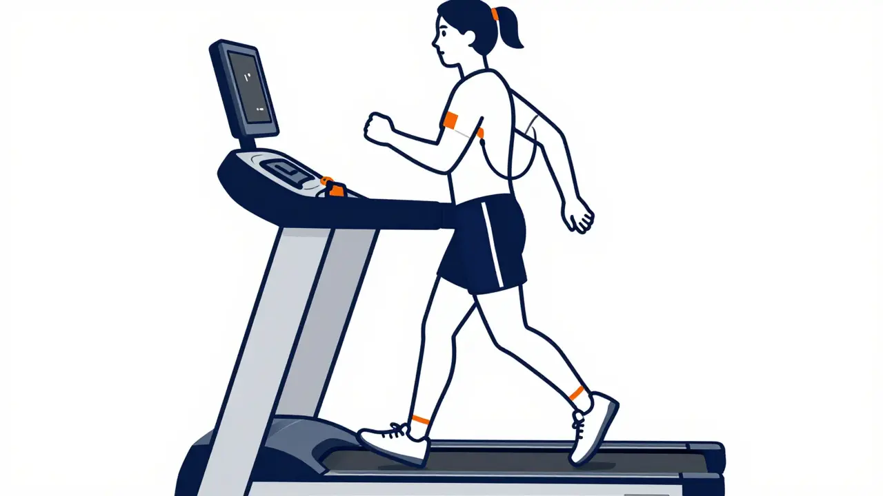
If a resting ECG is a snapshot, a Stress Test is a movie. It forces your heart to work harder, which reveals if there's a bottleneck in your blood flow. This is primarily used to diagnose Coronary Artery Disease (CAD), where the arteries supplying the heart muscle become narrowed.
For most people, this involves the Bruce protocol. You start on a treadmill at a slow pace (1.7 mph) with a slight incline. Every three minutes, the speed and the slope increase. The goal is to reach 85% of your maximum predicted heart rate. Why do this? Because when the heart muscle is stressed, any lack of oxygen (ischemia) causes the electrical waveforms on the ECG to change, specifically through ST-segment depression. Interestingly, the longer you can keep going, the better the prognosis; each extra minute of exercise is linked to a 12% drop in the risk of future cardiac events.
When You Can't Exercise: Chemical Stress Tests
Not everyone can jog on a treadmill. If you have severe arthritis, asthma, or are recovering from surgery, doctors use a pharmacological approach. Instead of a treadmill, they use drugs like Adenosine or Dobutamine to mimic the effects of exercise by dilating blood vessels or increasing the heart rate.
These tests take longer-usually 30 to 60 minutes-and can feel a bit more intense. Some patients report feeling shortness of breath or a strange pressure in the chest during the infusion. While it's uncomfortable, it's a safe way to get the same critical data as an exercise test without the physical strain.
Adding a Visual Layer: Echo and Nuclear Imaging
A standard ECG stress test only looks at electrical signals. To get a clearer picture, doctors often add imaging. Stress Echocardiography combines the stress test with ultrasound. This allows the cardiologist to actually see the heart walls moving. If a section of the heart stops pumping effectively during stress, it's a huge red flag for a blockage in that specific artery, with an accuracy rate of about 92%.
Then there's Nuclear Stress Testing. This involves injecting a radioactive tracer like Technetium-99m. As the tracer flows through your blood, a special camera tracks where it goes. If the tracer doesn't reach a certain part of the heart during exercise, it confirms a lack of blood flow. While it's highly sensitive (around 85-89%), it does expose you to radiation, which is why ultrasound-based tests are often preferred for lower-risk patients.
Gender Gaps and Diagnostic Accuracy
One critical thing to keep in mind is that these tests aren't perfect for everyone. There is a known issue with false negatives in women, especially premenopausal women. This happens because women's heart physiology and microvascular functions differ from men's. A standard stress ECG might look "normal" even when there is an underlying problem.
To fix this, newer techniques like speckle-tracking strain analysis are being used with echocardiography. This advanced imaging increases the sensitivity for detecting microvascular dysfunction in women from 68% up to 89%. If you're a woman experiencing symptoms but getting "clear" results on a standard stress test, it's worth asking your doctor about these more detailed imaging options.
What to Expect and How to Prepare
Preparation is key to getting an accurate result. The biggest culprit for a ruined test is caffeine. You'll likely be asked to avoid coffee, tea, and soda for 24 hours before a stress test. Why? Because caffeine can interfere with the pharmacological agents used in chemical tests and can artificially spike your heart rate.
On the day of the test, wear comfortable clothes and sneakers. You'll be monitored for four main things: your heart rate, your blood pressure, the ECG waveforms, and your oxygen levels. Don't be surprised if the technician has to pause to fix an electrode-movement during a treadmill test often causes "artifacts" or static on the reading, which happens in about 18% of tests.
Is a stress test dangerous?
For most people, it is very safe because you are monitored by medical professionals in a controlled environment. However, it is generally avoided if you've had a heart attack in the last two days or if you have unstable heart rhythms. Always disclose your full medical history to your doctor beforehand.
How long does it take to get the results?
In about 85% of facilities, a technician or doctor can provide a preliminary reading immediately after the test. A full, detailed report usually takes a bit longer as the cardiologist reviews the waveforms and images carefully.
What is the difference between an EKG and an ECG?
There is no difference. ECG stands for electrocardiogram, and EKG comes from the German spelling "Elektrokardiogramm." They are exactly the same test.
What does a "positive" stress test mean?
In medical terms, a "positive" result means the test found evidence of the problem it was looking for-usually that your heart isn't getting enough oxygen during exertion, which suggests potential coronary artery disease.
Can a stress test detect a heart attack?
A resting ECG is better for detecting an active or past heart attack. A stress test is designed to find the *risk* of a heart attack by identifying blockages before they cause a full event.
Next Steps After Your Results
If your test comes back normal, it's usually a great sign, but keep in mind that no single test is 100% foolproof. If you're still feeling chest pain or shortness of breath, don't ignore it just because the ECG was clear.
If the result is "positive" or inconclusive, your doctor might suggest a Coronary CT Angiography (CCTA). This is a more detailed scan that looks at the actual structure of the arteries rather than just the function. Depending on your risk level, the next step could also be a cardiac catheterization, where a tiny tube is used to inject dye directly into the heart to find the exact location of a blockage.

15 Comments
The distinction between resting electrical activity and stress-induced ischemia is fundamental for an accurate diagnosis of coronary artery disease.
Absolute game changer for the ladies! It's wild how often we're just told 'everything is normal' while feeling like our hearts are doing gymnastics in our chests. Seriously, push for that speckle-tracking if you're not feeling the standard results. Don't let them brush you off!
One finds it quaint that the general public is just now discovering the gender disparity in cardiac diagnostics. It is a rather pedestrian observation, yet the lack of widespread implementation of advanced imaging suggests a systemic intellectual lethargy within the medical establishment. Truly, the gap between theoretical knowledge and clinical application is an abyss.
Wait a minute... radioactive tracers? They're literally pumping glowing chemicals into our veins and then using a 'special camera' to see where it goes? Sounds like some kind of government tagging system to me. Why the secrecy about what's actually in those dyes? Total madness!
This is so helpful!! I had a stress test last year and I was so nervus because the tread mill kept speeding up and I thought I was gonna fall off lol. The doctor told me about the ST-segment but I didn't realy get it until now. Thank you for making it simple to understand for us regular peeple!!
The sheer inefficiency of the Bruce protocol is staggering. To suggest that a linear increase in speed and incline is an optimal measure of cardiovascular stress is a laughable simplification of human physiology. It is an archaic methodology that persists only through the stubborn inertia of medical tradition, which is utterly pathetic in an era of precision medicine.
I appreciate the explanation regarding the chemical alternatives for those with mobility issues.
Omigosh, the caffeine thing is such a bummer!
I live for my morning brew, and imagining a whole 24 hours without a single sip of coffee is practically a nightmare. But hey, better to be a bit grumpy than to have a wonky test result, right? Totally worth it to get the real deal on heart health!
Honestly, I only ever go to the best clinics in the city because they actually use the high-end imaging mentioned here. Why settle for a basic treadmill when you can have the gold standard? It's just a matter of knowing which doctors are actually in the loop and which ones are still living in the 90s. Stay fancy, everyone!
The point about false negatives in women is a crucial takeaway here. We need to be more assertive in asking for the advanced imaging if the baseline tests don't align with our actual physical symptoms. It's about taking charge of our own health outcomes.
I agree, it's so important to advocate for yourself!
The ST-segment depression is a key marker for myocardial ischemia. The hemodynamic response during peak exertion provides a critical window into the perfusion limits of the coronary arteries. This helps in calculating the functional capacity and risk stratification for the patient.
Life is but a series of rhythms... and some are just slightly off-beat!!!
It is truly a blessing that science can peek inside our bodies to keep us safe. We should all be grateful for these tools that allow us to live longer and healthier lives together. Wishing everyone peace and strong hearts! :) 💖
American medicine is the best. Period.